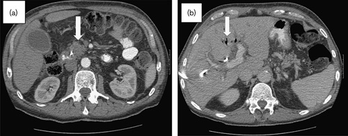
Fig. 7.

February 2011 presentation to the emergency room, where the patient was found to be in septic shock 2 weeks after biliary stent placement. (a) Contrast-enhanced CT scan: stable size of mass in the uncinate process of pancreas (arrow) and no apparent tumor progression. There was mild diffuse induration of the mesentery consistent with third spacing of fluid. (b) Contrast-enhanced CT scan: hepatic parenchyma with air in the biliary tree (arrow), but no parenchymal lesions identified. CT, computed tomography.
