
Fig. 5.
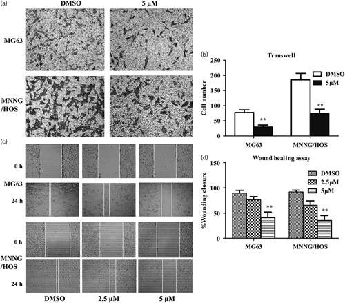
(S)-Crizotinib inhibited the migratory activities of osteosarcoma cells. (a) Transwell assay of osteosarcoma cells treated with DMSO or 5 μmol/l (S)-crizotinib: images were taken at magnification ×100. Photos are representative fields of cells penetrating the membrane and fixed in pure methanol, stained with crystal violet, and counted. the membrane. (b) Quantitative analysis of the transwell assay. Bars are means±SD from three independent experiments. **P<0.01 compared with the DMSO group, Student’s t-test. (c) Wound-healing assay of osteosarcoma cells treated with DMSO, 2.5, or 5 μmol/l (S)-crizotinib: images were taken at magnification ×40. (d) Quantitative analysis of the wound-healing assay. The percentage of wound closure=original width–width of 24 h after cell migration/original width. Bars are mean±SD from three independent experiments. **P<0.01 compared with the DMSO group, one-way analysis of variance, followed by the least significant difference test.