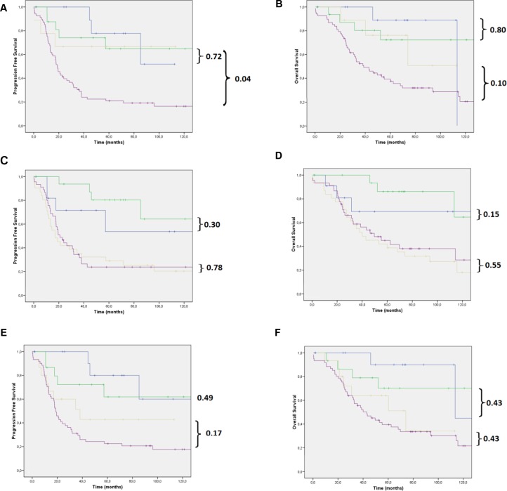
Figure 2.
(A) PFS and (B) OS of women with LGSOC and HGSOC in initial and advanced disease stages based on morphological differentiation (WHO); (C) PFS and (D) OS of women with LGSOC and HGSOC in cases of negative and positive WT1 expression; (E) PFS and (F) OS of women with LGSOC and HGSOC in initial and advanced disease stages based on the immunohistochemical p53/p16 algorithm. All our analyses were performed in patients with stage I + II (blue and green lines) or in patients with stage III + IV (yellow and purple lines) disease. Note: Blue line: LGSOC stage I + II; green line: HGSOC stage I + II; yellow line: LGSOC stage III + IV; purple line: HGSOC stage III + IV.

