Figure 1.

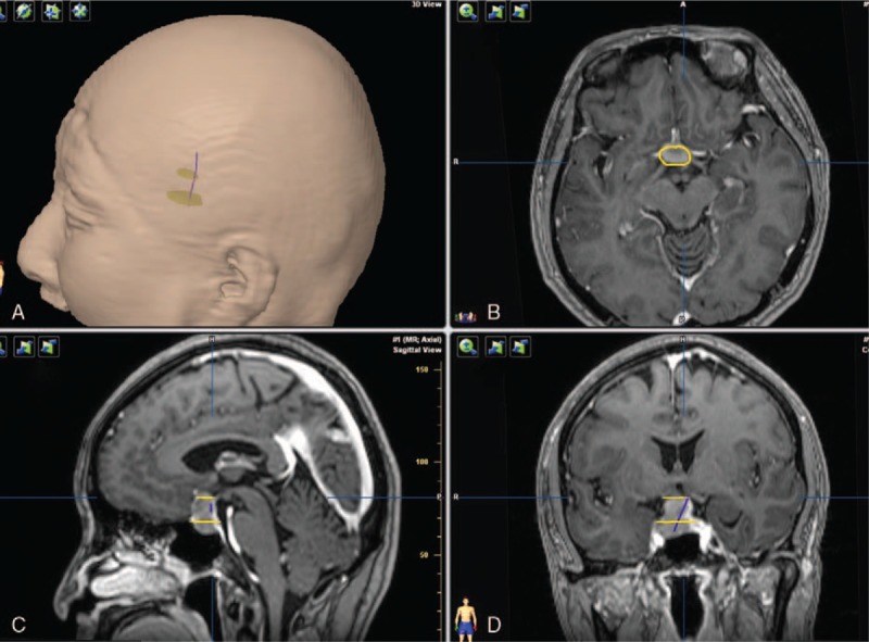
HHT visualization in a patient with pituitary adenoma. (A) The contour of head with 2 ROIs and HHT. (B) The yellow contour of adenoma in axial plane. (C) Two yellow plane show 2 ROIs masks. (D) HHT after proper pruning.

HHT visualization in a patient with pituitary adenoma. (A) The contour of head with 2 ROIs and HHT. (B) The yellow contour of adenoma in axial plane. (C) Two yellow plane show 2 ROIs masks. (D) HHT after proper pruning.