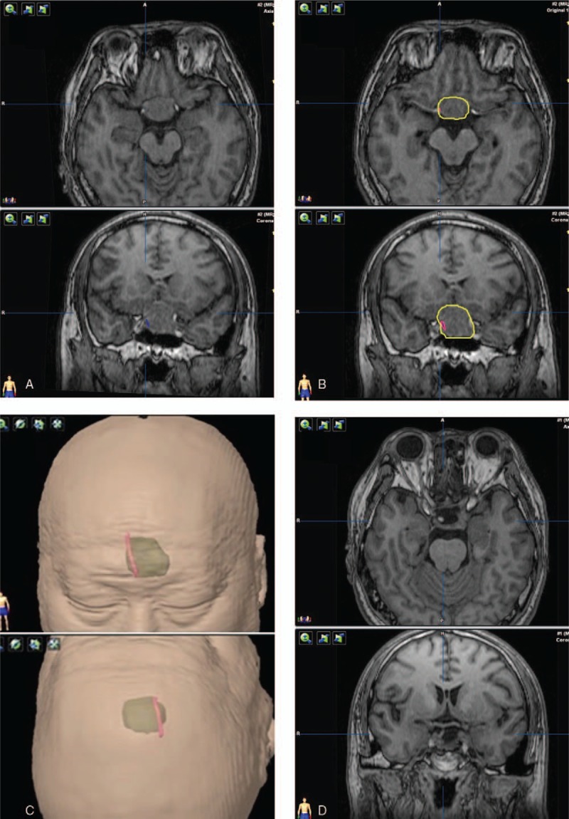
Figure 4.

HHT visualization and the postoperative observations of pituitary stalk. The tracts and pituitary stalk are located in the right portion of the tumor. (A) Tracts (blue fibers) visualization with the adenoma. (B) Red contour of the tracts and yellow contour of the adenoma. (C) The green translucent part shows a 3D anatomical model of the tumor, and red part shows the contour of tracts. (D) The image of postoperative pituitary stalk.
