Figure 3.

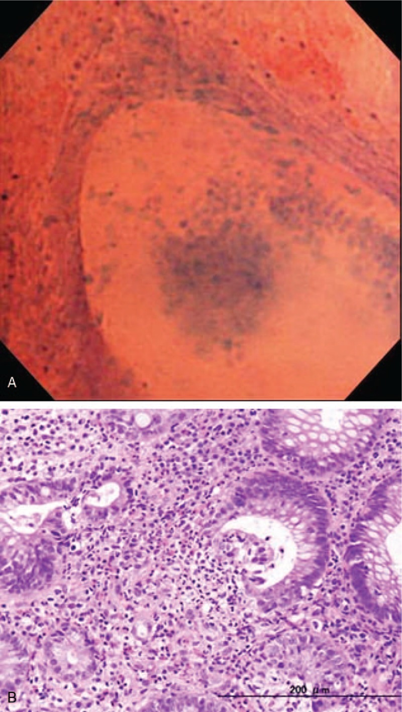
EC shows the irregularly dilated pits and intraglandular infiltration of inflammatory cell components (A). Histopathology using the targeted biopsy specimen shows crypt abscess formation within the dilated crypt (B).

EC shows the irregularly dilated pits and intraglandular infiltration of inflammatory cell components (A). Histopathology using the targeted biopsy specimen shows crypt abscess formation within the dilated crypt (B).