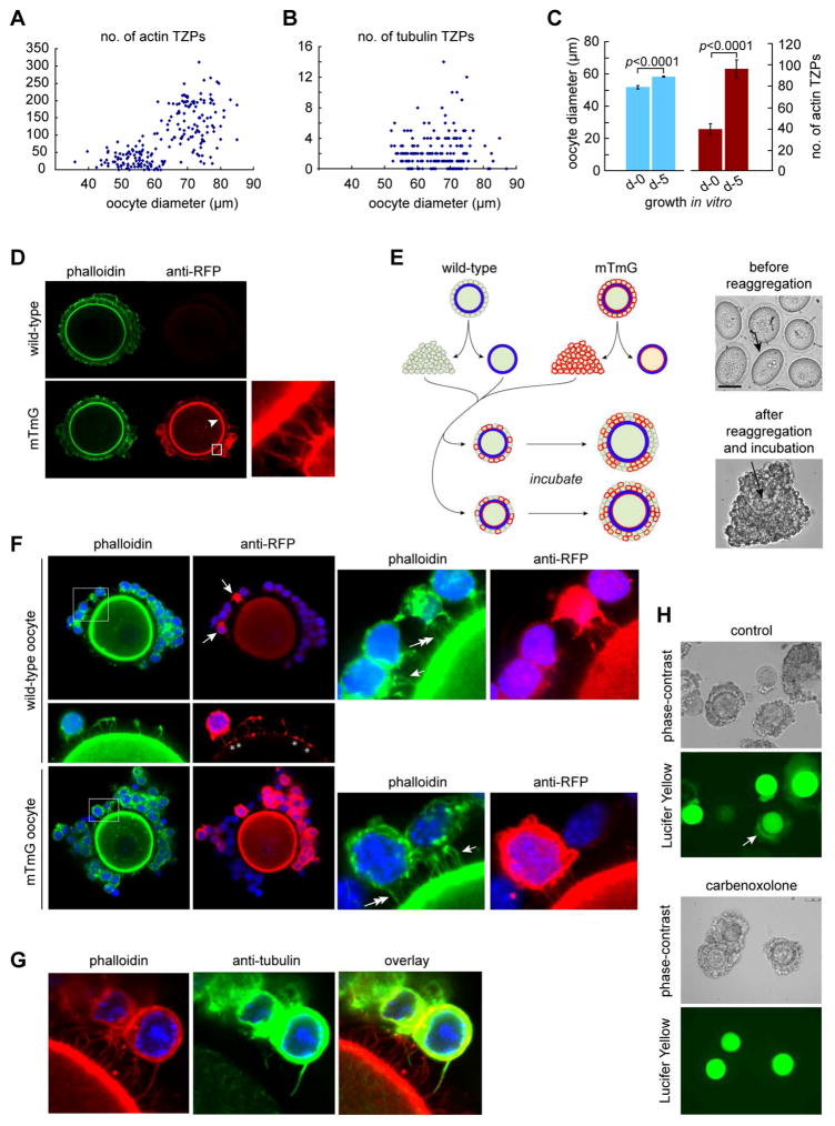
Figure 2. Granulosa cells elaborate new TZPs during oocyte growth.
(A) Oocytes at different stages of growth were stained using phalloidin and the number of TZPs in an equatorial confocal optical section was counted. (B) As in (A) except that the oocytes were stained using anti-tubulin. (C) Oocyte diameter and number of actin-TZPs were determined in GOCs immediately after isolation or after 5 days of growth in vitro. (D) GOCs of wild-type (upper) or mTmG (lower) mice were stained using phalloidin and anti-RFP. Anti-RFP stains TZPs (inset) as well as the oocyte membrane (arrowhead) of mTmG mice. (E) Left: Reaggregation strategy. Right, upper: oocytes prior to reaggregation. Arrow indicates zona pellucida. Right, lower: reaggregated complex after 5 days of incubation. Arrow indicates oocyte. (F) Reaggregated complexes after incubation. Some granulosa cells have been removed to enable TZPs to be seen more clearly. Upper: Wild-type oocyte, whose membrane is not stained by anti-RFP, enclosed by wild-type and mTmG (arrows) granulosa cells. Right panel shows enlargement of the boxed area. TZPs of wild-type granulosa cells are stained by phalloidin only (single arrow); TZPs of mTmG granulosa cells are also stained by anti-RFP (double arrow). Middle: Asterisks illustrate where TZPs of mTmG granulosa cells contact the surface of a wild-type oocyte. Lower: mTmG oocyte, whose membrane is stained by anti-RFP, enclosed by wild-type and mTmG granulosa cells. Right panel shows enlargement of the boxed area. Single arrow and double arrows indicate TZPs as above. (G) TZP stained by both phalloidin and anti-tubulin. (H) Lucifer Yellow was injected into the oocyte of reaggregated complexes. Upper: bright-field; lower: dark-field. Arrow shows fluorescence in granulosa cells. Lower paired panels show complexes incubated in the gap junction blocker, carbenoxolone. See also Figure S1.

