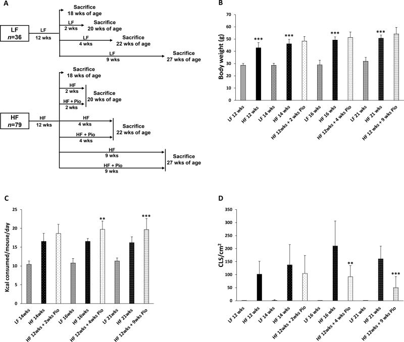
Figure 3. Treatment with pioglitazone led to time-dependent suppression of periprostatic white adipose tissue inflammation.
A, study schema. At 6 weeks of age, C57BL/6J male mice were fed either a low fat (LF) or high fat (HF) diet. A group of LF diet fed mice was sacrificed at 18 weeks of age after 12 weeks on LF diet. A second group of LF diet fed mice was sacrificed at 20 weeks of age after 14 weeks on LF diet. A third group of LF diet fed mice was sacrificed at 22 weeks of age after 16 weeks on LF diet. The fourth group of LF diet fed mice was sacrificed at 27 weeks of age after 21 weeks on LF diet. The HF diet fed mice were randomized to seven groups at 18 weeks of age after 12 weeks on HF diet. One group was sacrificed at 18 weeks of age while three other groups were continued on HF diet for an additional 2, 4 or 9 weeks. The remaining three groups were switched to HF diet containing 0.06% w/w pioglitazone for an additional 2, 4 or 9 weeks. B, body weights of mice in different treatment groups. C, caloric consumption was monitored in mice following initiation of pioglitazone treatment. Average calorie consumption during the treatment period is presented. D, periprostatic white adipose inflammation was quantified as CLS/cm2. B–D, mean ± SD (error bars) are shown. B, n=6–16/group, ***p<0.001 compared with LF diet group. C, n=6–16/group; **p<0.01 compared with HF diet group. D, n=6–12/group; **p<0.01, ***p<0.001 compared with HF diet group.

