Fig. 2.

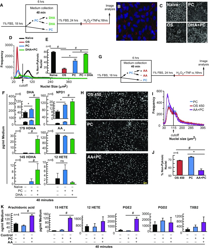
DHA enhances and AA attenuates PC in human RPE cells in vitro. APRE-19 and human primary cells were treated with DHA, AA, or vehicle 6 h prior to mild oxidative preconditioning and lethal OS. Medium was collected for lipid analysis and nuclei were imaged for cell survival using MAIM. a Experimental design illustration for DHA using primary human RPE cells. The following are examples of the characterization of these cell responses: b Confocal image 20× magnification and zoomed-in human primary RPE cells stained with Hoestch 33258 and probed for ZO-1; c 8-bit grey-scale confocal image (20× magnification) of human primary RPE cells stained with Hoestch 33258; and d nuclear size frequency histogram plots for PC + DHA. e Quantification of frequency plots as a percentage of non-nuclear pyknosis (% survival) in cells preconditioned with ±50 µM H2O2 and/or DHA (200 nM) for 6 h prior to lethal OS challenge. f UPLC-ESI-MS/MS quantification of AA, DHA, and derivatives 12-HETE, 17-HDHA, 14-HDHA, and NPD1 at 40 min PC and PC + 200 nM DHA. g Experimental design for AA and ARPE-19 cells. The following are examples of the characterization of these cell responses: h 8-bit grey-scale confocal image (20× magnification) of ARPE-19 cells stained with Hoestch 33258; and i nuclear size frequency histogram plot for PC + AA. j Quantification of frequency plots as a percentage of non-nuclear pyknosis (% survival) in cells preconditioned with ±50 µM H2O2 and/or 200 nM AA for 6 h prior to lethal OS challenge. k UPLC-ESI-MS/MS quantification of AA and derivatives 15-HETE, 12-HETE, PGE2, PGD2 and TXB2 at 40 min PC and PC + 200 nM AA. All lipid concentrations were normalized using deuterium-labeled internal standards and to medium volume. *p < 0.05, # p < 0.001, scale bar 10 µM
