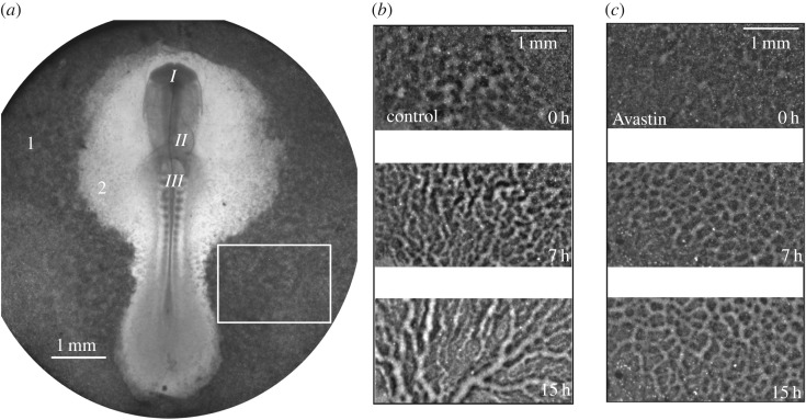
Figure 1.
(a) Whole embryo image. From top to bottom, around the middle line, we see the head fold I, forming heart II and somites III, which are blocks of cells which will give rise to muscles, bone, cartilage and skin. The number 1 indicates the area opaca and 2 is the area pellucida. A selected rectangle (6.58 mm2) in the area opaca is used to monitor the forming vascular network. A zoom of the rectangular area was taken from (b) a control embryo, (c) an Avastin-treated embryo, at times: 0, 7 and 15 h, from top to bottom.

