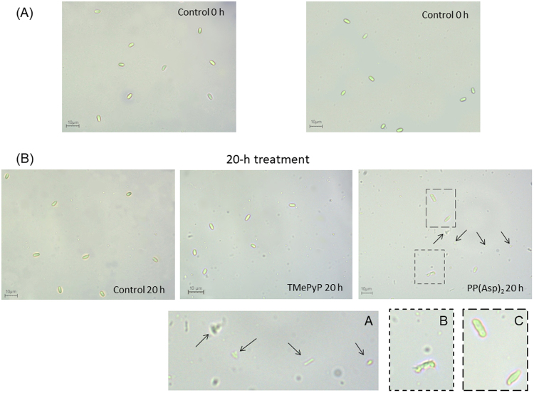
Figure 8.
Light microscopic images of the microsporidia showing morphological differences between porphyrin-treated and untreated spore preparations. (A) At the beginning of incubation with porphyrins and (B) after 20 h of treatment. Morphological changes and cell debris are marked with dashed outlines and arrows (60× objective). Insets showing higher magnifications (dashed lines) are shown in panels (A,B) and (C). Spores with deformed walls are visible in panel (C).

