Figure 5.
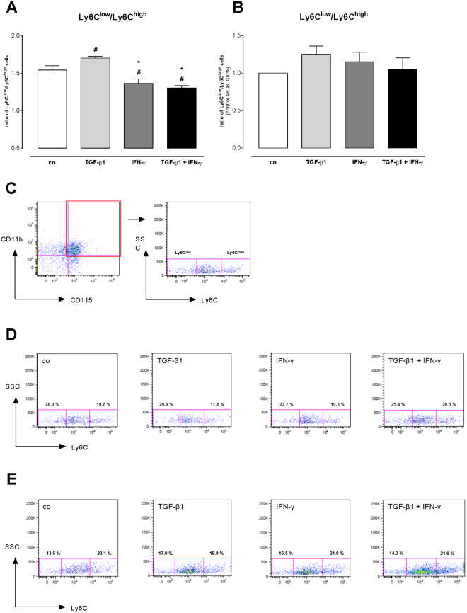
IFN-γ- and TGF-ß1-induced monocyte migration. (A) Flow cytometry analysis of CD11b+CD115+Ly6C+ stained cells shows differences between the ratios of Ly6Clow/Ly6Chigh monocytes versus the different conditioned media in C4 fibroblasts. (B) Indication for enhanced migration of Ly6Clow monocytes towards TGF-ß1-conditioned media of cardiac fibroblasts, whereas co-stimulation with TGF-β1 + IFN-γ increases the number of migrated Ly6Chigh monocytes. (C) Gating strategy to define the amount of CD11b+CD115+Ly6C+ cells. (D) Representative dot blots for the migrated monocytes of the different conditioned media of stimulated C4 fibroblasts (n = 6/condition). (E) Representative dot blots for the migrated monocytes of the different conditioned media of stimulated cardiac fibroblasts (n = 3/condition). Data are shown as ratio of Ly6Clow/Ly6Chigh monocytes of 3 independent experiments. Statistical analysis was performed by One way-ANOVA or Kruskal Wallis test (#p < 0.05 vs co, *p < 0.05 vs TGF-β1, §p < 0.05 vs IFN-γ).

