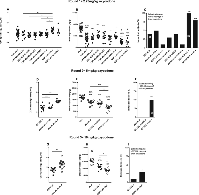
Figure 1.
Modulation of IL-4 signaling enhances anti-oxycodone antibody, vaccine efficacy against oxycodone, and the subset of immunized subjects that showed vaccine efficacy. Three independent cohorts of male BALB/c mice were immunized s.c. with OXY-KLH and KLH adsorbed on alum adjuvant and challenged a week after the 3rd immunization with increasing doses of oxycodone (2.25–10 mg/kg s.c.). Interleukins were administered at days 0, 14 and 28 in combination with each immunization. The mAb was administered 2 days prior and 1 day after the 1st immunization. Experimental groups: (A–C) KLH (n = 24), OXY-KLH (n = 25), and OXY-KLH plus: rIL-4 (30,000 IU, s.c.), rIL-4 (60,000 IU, s.c.), IL-4 plus IL-21 (60,000 IU total, s.c.), αIL-2 alpha receptor mAb (αCD25, 1.0 mg, i.p., n = 10), αCD25 mAb (1.0 mg, i.p.) plus IL-4 (30,000 IU, s.c., n = 10), αIL-4 mAb (1.0 mg, i.p., n = 10), or IL-4:αIL-4 mAb (30,000 IU and 0.5 mg of mAb mixed prior to injection, s.c., n = 5), and challenged with 2.25 mg/kg oxycodone. (D–F) KLH, OXY-KLH, and OXY-KLH plus αCD25 mAb (1.0 mg, i.p.), or αIL-4 mAb (1.0 mg, i.p.) and challenged with 5 mg/kg oxycodone (n = 10/group). (G–I) KLH, OXY-KLH, and OXY-KLH plus αIL-4 mAb (1.0 mg, i.p.) and challenged with 10 mg/kg oxycodone (n = 10/group). Shown oxycodone-specific IgG titers, oxycodone distribution to the brain, and fraction (%) of immunized subjects that showed more than 50% reduction in oxycodone distribution to the brain compared to the mean brain concentration in the KLH control group. Data are mean ± SEM. Data shown are from at least two independent experiments. Titers and oxycodone concentrations analyzed by one-way ANOVA and Tukey’s multiple comparisons test or unpaired two-tailed t-test. Chi-square test: (C) (χ2 = 282.8, df = 8), F) (χ2 = 120.0, df = 2), and I) (χ2 = 12.5, df = 1). Percentages (%) above bars indicate reduction compared to KLH. *p < 0.05, **p < 0.001, ****p < 0.0001 compared to KLH, and brackets or # indicate significance between groups, #p < 0.05.

