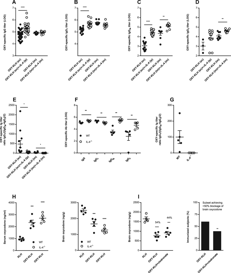
Figure 3.
Depletion and ablation of IL-4 shifts post-vaccination IgG subclass distribution. (A–E) Serum anti-oxycodone IgG antibodies were analyzed for IgG1, IgG2a, and IgG3 subclass in mice immunized with OXY-KLH ± αIL-4 mAb from experiments shown in Figs 1A–H and 2A–C. (A) oxycodone-specific IgG titers in mice immunized with OXY-KLH s.c., n = 49; OXY-KLH s.c. + IL-4 mAb i.p., n = 38; OXY-KLH i.m., n = 8; OXY-KLH i.m. + IL-4 mAb i.m., n = 8). IgG subclass analysis was performed on a randomly selected sample from each immunization group (n = 5–6 mice/group): (B) IgG1, (C) IgG2a, (D) IgG3, (E) IgG1/((IgG2a + IgG3)/2). (F–H) In a separate study, IL-4−/− and wild-type mice controls were immunized with KLH or OXY-KLH in alum (s.c., n = 5 each group). (F) oxycodone-specific IgG titers by subclass, (G) IgG1/((IgG2a + IgG3)/2), and (H) effect of immunization on oxycodone distribution to serum and brain. (I) In a follow-up independent experiment, mice were immunized with either KLH (s.c., n = 4) or OXY-KLH (s.c., n = 10). The OXY-KLH group received either saline or liposome-encapsulated clodronate to deplete macrophages (n = 5), and 24 hrs later challenged with 5.0 mg/kg oxycodone. Data are from one experiment. (I) vaccine efficacy in blocking oxycodone distribution to the brain (left) and the subset (%) of immunized mice showing efficacy (right). Data are mean ± SEM. (A–G) unpaired two-tailed t-test. (H,I) One-way ANOVA paired with Tukey’s test, and chi-square test (χ2 = 8.00, df = 1, p = 0.0047). *p < 0.05, **p < 0.01, ***p < 0.001, ****p < 0.0001.

