Figure 1.
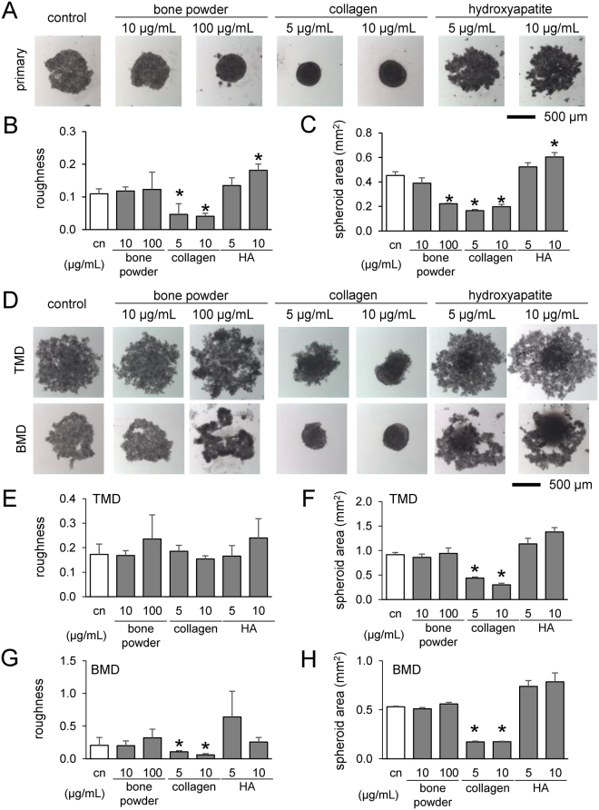
Formation of tumor spheroids in the presence of powdered bone extract (10 and 100 µg/mL), collagen (5 and 10 µg/mL), and hydroxyapatite (5 and 10 µg/mL) after 24 h. An asterisk (*) denotes p < 0.05 compared with control. (A) Tumor spheroids with primary breast cancer cells. (B & C) Roughness and cross-sectional area of primary cell spheroids, respectively. (D) Tumor spheroids with TMD and BMD cells. (E & F) Roughness and area of TMD cell spheroids. (G & H) Roughness and cross-sectional area of BMD cell spheroids.

