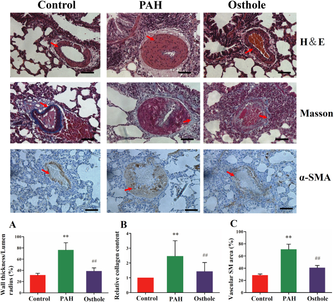
Figure 3.
Systems influence of osthole on pulmonary vascular remodeling. (A) H& E staining of rat lungs demonstrated that wall thickness was increased in the rats with PAH compared with healthy-control rats. (B) Deposition of collagen was obviously higher in the rats with PAH than that in healthy ones which was characterized by Masson staining. (C) Expression of α-SM actin was also markedly increased in the rats with PAH. These differences between PAH and control groups were considerably restored by osthole treatment (80 mg/kg/d). Data shows quantitative analyses of positive staining per vascular area. The data was denoted as mean ± S.E.M. (p value <0.05 was considered as statistically different between two groups (∗ or #), and p value < 0.01 (∗∗ or ##) was regarded as significantly different between two groups; *, ** was indicated to compare with control group; #, ## was designed to compare with PAH group).

