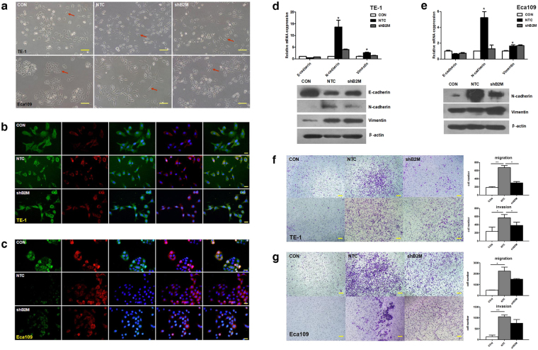
Figure 2.
MSCs-derived B2M promotes epithelial-mesenchymal transition and enhances mobility of ESCC cells in vitro. (a) Morphological appearance of esophageal cancer cells treated with conditioned medium of MSCNTC and MSCshB2M suggested MSCs-derived B2M promotes EMT in vitro. In mesenchymal status, TE-1 present morphological changes from cobblestone-like to cord-like cells with sharp protuberance while Eca109 present incompact structure instead of clustered ‘islets’. Red arrows indicate representative morphological changes of tumor cells. (b,c) Immunofluorescence imaging revealed increased expression of N-cadherin (red) accompanied with decreased expression of E-cadherin (green) after treatments with MSCNTC-CM but not with MSCshB2M-CM in (b) TE-1 and (c) Eca109 cells. Nucleuses were immunostained with DAPI (blue). From left to right the images show the staining results for E-cadherin, N-cadherin, E-cadherin + DAPI, N-cadherin + DAPI, and Merge. (d,e) The qRT-PCR (upper panel) and western blot (lower panel) results showed elevated N-cadherin and vimentin with down-regulated E-cadherin in TE-1(d) and Eca109 (e) cancer cells after treated with MSCNTC-CM in contrast to the MSCshB2M-CM. (f) The number of the TE-1 cells in the migration (upper panel) and the invasion (lower panel) assays after treatment with MSCNTC-CM was significantly increased compared with the negative control, while RNAi against B2M (MSCshB2M) partially blocked this effect. (g) Similar migration and invasion results were observed in the Eca109 cells. Data is expressed as mean ± S.D. of three independent experiments. Error bars indicate S.D. (n = 3); *p < 0.05, **p < 0.01; CON: tumor cells untreated with conditioned medium; Scales bars: 100 μm.

