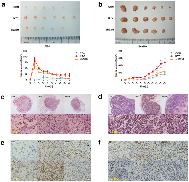
Figure 5.
MSCs-derived B2M enhances tumor development in vivo. (a,b) Representative macroscopical images of fresh BALB/c nude mice tumor tissues (upper panels) and the dynamic tumor growth curves in the TE-1 (a) and the Eca109 (b) cell group (lower panels). MSCNTC contributed to the enhancement of the xenograft tumor formation rate of the ESCC cells compared with the negative control, and knockdown of B2M reversed this tendency. The average tumor volume in the MSCNTC group was significantly larger than in mice injected with ESCC cells only, while the tumor volume in the MSCshB2M group was much smaller than that in the MSCNTC group until the sacrifice of animals. (c, d) H&E staining results showed the pathological structure of xenograft tumor section formed by TE-1 (c) and Eca109 (d) cells, including representative image of intact section (upper panels) and regional details (lower panels). (e,f) Immunohistochemical staining of TE-1 (e) and Eca109 (f) cells from the xenograft tumor tissue was performed using anti-PCNA (upper panel) and anti-Ki67 (lower panel) polyclonal antibodies. Tissue sections of MSCNTC group displayed more positive signals compared with the negative control and the MSCshB2M group. Data were expressed as mean ± SEM of three independent experiments. Error bars indicate SEM (n = 6); *p < 0.05; CON: mice transplanted with tumor cells alone; Scale bars: 100 μm.

