
Fig. 5.
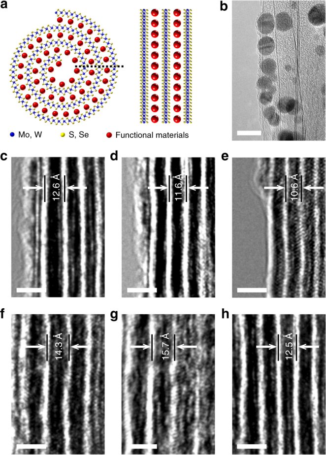
Hybrid transition metal dichalcogenide (TMD)-nanoscrolls (NSs). a Schematic showing the cross section and sidewalls of hybrid TMD-NSs. b–h HR-TEM images of sidewalls of MoS2-NSs hybridized with AuNPs (b), GO (c), pentacene (d), CuPc (e), PDPP3T (f), DNA (g) and polypeptide (h). AuNPs approximately 5 nm in diameter were clearly observed between the walls; the GO flakes were also distinguishable, as shown in c. Lattice expansion was displayed in all hybrid NSs above. The bright white walls represent the MoS2 layers in (c–h) (scale bars, 10 nm in b and 2 nm in c–h)