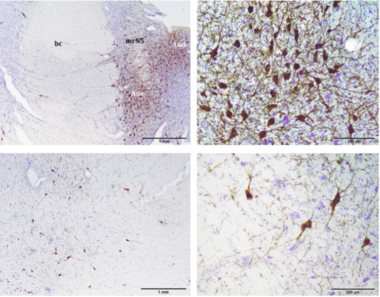
Figure 2.
A6d and A6v subdivisions (upper pictures) and A7 division (lower pictures). A6d is entirely restricted to the PAG and medial to the bc, while A6v is located just outside the PAG, ventro-lateral to the A6d and medial to the bc. A6d and A6v are separated by bundles of nerve fibres belonging to mrN5. A7 is located in the pontine tegmentum, ventral to A6. Atlantic spotted dolphin, Stenella frontalis. TH free-floating immunolabelling, counterstained with thionine. bc, brachium conjunctivum; mrN5, mesencephalic root of the trigeminal nerve.

