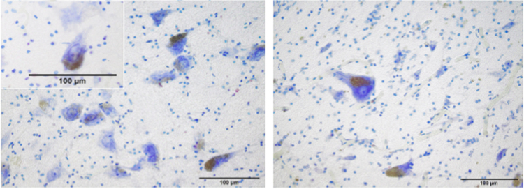
Figure 5.
Neuromelanin in the cytoplasm of the LC’ perikarya. Most neurons have the typical dark brown-blackish neuromelanin granules, occupying either a pole of the cell or even half of the cytoplasm. The nucleus is placed in a central position with an evident nucleolus. Around the nucleus, Nissl granules are distributed homogeneously. Atlantic spotted dolphin, Stenella frontalis (left) and Risso’s dolphin, Grampus griseus (right). Thionine staining.

