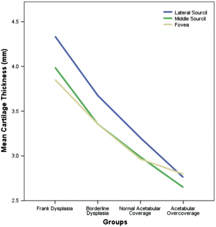
Fig. 3.
Cartilage thickness at the lateral sourcil (blue) was significantly increased in all clinical subgroups relative to that at the middle sourcil (green) or fovea (yellow), with the latter two measurements being statistically equivalent. Cartilage thickness at all measurement locations was inversely proportional to the degree of lateral acetabular coverage.

