
Fig. 1.
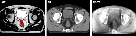
Transverse images of the lower abdomen of a rectal cancer patient using different imaging modalities. The images are of the same patient, depicting the same location through the rectal tumor. On the left; MRI, in the middle; computed tomography (CT) and at the right; CBCT. The tumor is delineated in red on the MR image. Note: on CT and CBCT images, there is contrast between the rectum and surrounding mesorectal fat, but no contrast between the rectum and the tumor