Fig. 2.

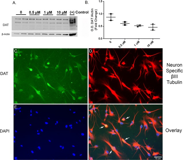
Human dopaminergic neurons express Dopamine Transporter. A. Representative western blot (A) and optical density (B) of dopamine transporter (DAT) show the presence of DAT in the lysates from vehicle and 27-OHC-treated neurons. Immunofluorescence imaging shows immunopositive staining for DAT in untreated neurons (C; green). Immunofluorescence for the neuron specific β-III Tubulin marker (D; red) and for nuclear counterstaining with DAPI (E; blue). F Overlay of dopamine transporter, neuron specific β-III Tubulin, and DAPI showing both nuclear and cytoplasmic localization of DAT (arrows)
