Fig. 6.

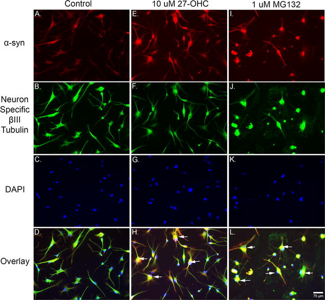
Both 27-OHC and the proteasomal inhibitor MG-132 increase α-syn protein levels. Immunofluorescene imaging shows that both 27-OHC (E) and MG132 (I) increase the immunostaining of α-syn compared to control untreated cells (A). Staining with the Neuron Specific βIII-Tubulin marker in control (B), 27-OHC-treated (F) and MG132-treated (J) neurons. Staining with the nuclear counterstain DAPI in control (C), 27-OHC-treated (G) and MG132-treated (K) neurons. The overlay shows multiple neurons exhibiting nuclear α-syn staining (arrows) in 27-OHC (H) and MG132 (L) treated neurons compared to untreated neurons (D)
