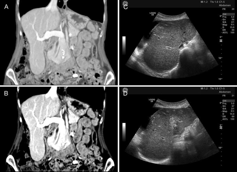
Fig. 1.

Findings on imaging. Computed tomography (CT) of the patient’s abdomen (A—abdominal view, and B—liver view) revealed a prominent Riedel lobe of the liver, with no evidence of obstruction, thrombosis, or malignancy. Mild changes of sigmoid diverticulosis were seen, with no evidence of acute diverticulitis. A repeat CT revealed mild ascites localized to the right upper quadrant, and the liver edge appeared to have a slightly nodular contour, a non-specific finding in this particular case, but which in general may suggest cirrhosis. Abdominal ultrasound (C, D) revealed a heterogeneous echotexture of the liver with regions of increased echogenicity in the right lobe, a non-specific finding which may be seen in hepatitis
