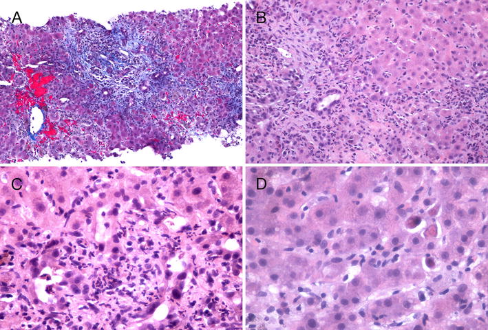
Fig. 2.

Findings on liver core biopsy. Ultrasound-guided core needle biopsy of the liver demonstrated that the overall hepatic architecture was preserved. Abnormal features included the marked expansion and fibrosis of portal tracts. In A fibrous tongues can be seen extending outwards into the liver parenchyma. The centrilobular areas appear preserved. There is pericentral sinusoidal congestion. Within the portal tracts (B) there are heavy infiltration of acute and chronic inflammatory cells, mostly lymphocytes and neutrophils. This extends into the limiting plate and extensively involves the adjacent liver parenchyma. Many damaged bile ductules and necrotic hepatocytes can be seen in C, and isolated necrotic hepatocytes can also be seen in the liver parenchyma in D. These biopsy findings are consistent with severe subacute hepatitis with developing portal fibrosis. The presence of plasma cells within the inflammatory infiltrate suggests the likelihood of an immune mechanism (hypersensitivity) as a component of the damage. However, the overall pattern of liver damage is unlike typical autoimmune hepatitis. In the absence of anti-smooth muscle antibodies, the possibility of drug-induced hepatitis via a hypersensitivity or immune mechanism is favoured. In particular, bile ductular damage—which is commonly seen in drug-induced hepatitis—may in turn produce hypersensitivity-related liver damage
