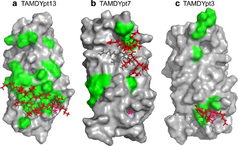
Fig. 5.

Typical peptide conformation. Typical conformations observed along TAMD trajectories TAMDYpt13 (a), TAMDYpt7 (b) and TAMDYpt3 (c), for the peptide and the YEATS domain. The surface of YEATS domain is shown, colored in grey, and the residues with atomic contacts larger than 50% (Table 4), are colored in green. The peptide conformation is displayed in sticks, colored in red and the position of the acK18 native binding site is marked with an asterisk
