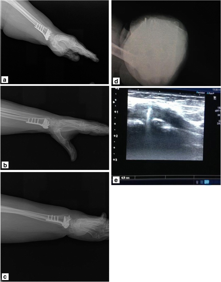
Fig. 1.

Patient number 8. Left wrist fracture, second compartment, 4.9 mm penetration. Postoperative lateral, 45° supination, 45° pronation, dorsal tangential and USG images. a Lateral. b Penetrating screw was not detectable on the 45° pronation radiograph. c Penetration was barely seen on the 45° supination radiograph. d Screw penetration on tangential radiograph. e 4.9 mm penetrating screw in the second compartment was seen by ultrasonography
