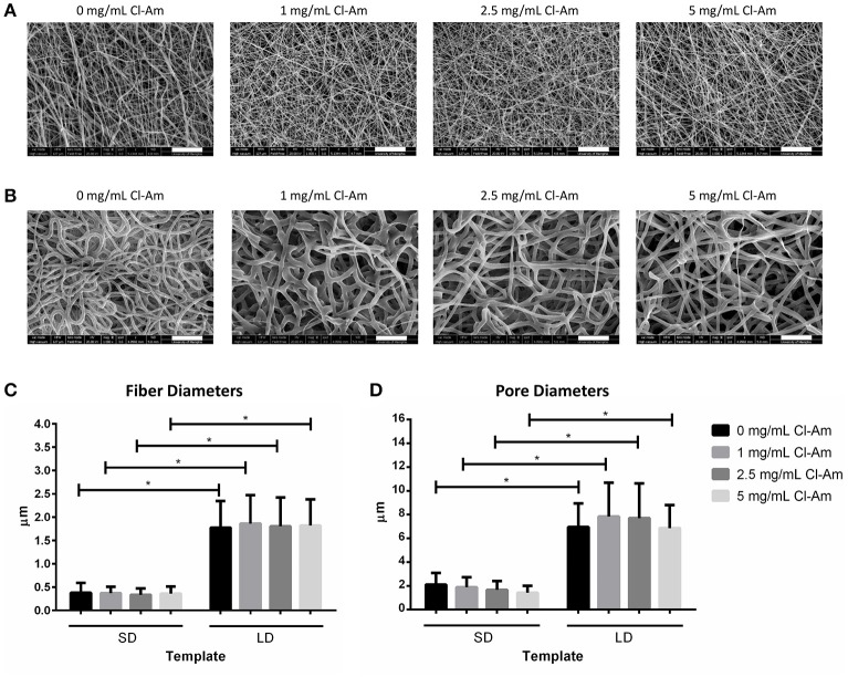
Figure 3.
PDO electrospins at low and high concentrations with Cl-Am. Representative SEMs of (A) SD and (B) LD templates with 0–5 mg/mL Cl-Am (scale bars = 20 μm). For all drug concentrations, all SD and all LD templates have statistically equivalent (C) fiber and (D) pore diameters; however, at each drug concentration, SD templates have significantly smaller (C) fiber and (D) pore diameters than the large diameter templates. *Significant difference (p < 0.05). (mean ± std. dev., n > 250 for fiber diameter analysis and n = 60 for pore diameter analysis).

