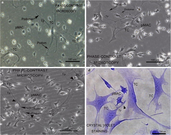
Fig. 2.

Real-time dynamic observation of primary directly co-cultured TCs and pMACs at 12 h intervals under phase-contrast microscopy; mice uterus. TCs have small oval cell bodies and long Tp branched from the cell body, appearing as an alteration of podom and podomer. a At 0 h, pMACs showed its normal regular round shape and no signs of activation. b At 12 h, pMACs showed moderate activation with irregular polyhedron shape and pseudopodia, but without any intercellular contacts to TCs. c At 24 h, pMACs exhibited intensive morphological changes and continuous activation, with obvious polyhedron, abundant pseudopodia; more importantly, direct heterocellular junctions can be observed between the activated pMACs and TCs (black arrowhead). d At 24 h, vital staining with crystal violet demonstrated heterocellular junction between the pseudopodia of activated pMACs and TCs (black arrowhead)
