Fig. 3.

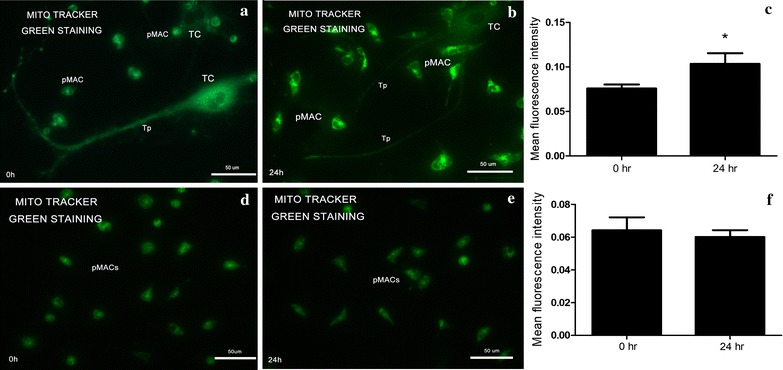
Fluorescence microscopy of Mito Tracker green staining. Error bars = SD. The MFI of each group was repeatedly detected three times under the same conditions. a–c The observed fluorescent intensity for TCs and pMACs in the co-cultured system at two time points. MFI at 24 h was significantly higher than that of 0 h (*P < 0.05), indicating active energy metabolism of pMACs in the co-cultured system. d–f No significant difference for background MFI of cultured-alone pMACs was found in two time points
