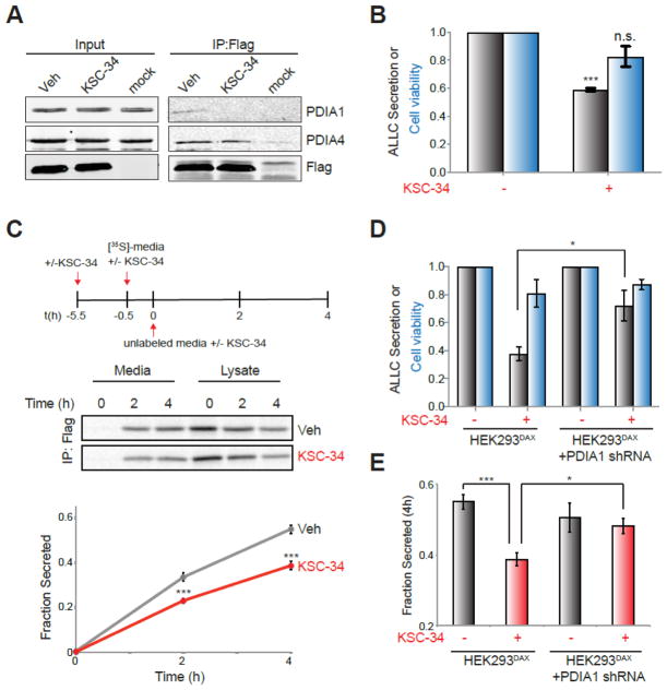
Figure 6.
KSC-34 reduces secretion of destabilized ALLC from mammalian cells. (A) Immunoblot of anti-FLAG IPs of lysates prepared from HEK293DAX cells transiently transfected with FTALLC and pre-treated for 1 h with vehicle or KSC-34 (40 μM). Cells were crosslinked for 30 min with DSP (500 μM) prior to lysis. Mock transfected cells are included as a control. (B) Graph showing secreted FTALLC (grey) and viability (blue) of HEK293Trex cells stably expressing FTALLC pretreated for 4 h with KSC-34 (40 μM). Media was conditioned for 2 h in the presence or absence of KSC-34 (40 μM) prior to quantification of secreted FTALLC by ELISA. Viability was measured following media conditioning by Cell Titre Glo. All data are normalized to vehicle-treated cells. Error bars show SEM for n=3 experiments. ***p<0.005. (C) Representative autoradiogram and quantification of the fraction [35S]-labeled FTALLC secreted from HEK293DAX cells using the experimental paradigm shown. Experiments were performed in the absence or presence of KSC-34 (40 μM) added 1 h prior to labeling and then again throughout the experiment. Fraction secreted was calculated as described in the Supporting Information 44. Error bars show SEM for n=4. ***p<0.005. (D) Graph showing secreted FTALLC (grey) and viability (blue) of HEK293DAX cells transiently expressing FTALLC pretreated for 4 h with KSC-34 (40 μM). HEK293DAX cells stably expressing PDIA1 shRNA are indicated. Media was conditioned for 2 h in the presence or absence of KSC-34 (40 μM) prior to quantification of secreted FTALLC by ELISA. Viability was measured following media conditioning by Cell Titre Glo. All data are normalized to vehicle-treated controls. Error bars show SEM for n=3 experiments. *p<0.05. (E) Graph showing the fraction [35S]-labeled FTALLC secreted at t=4 h from HEK293DAX cells transiently transfected with FTALLC quantified using the same experimental paradigm shown in Fig. 6C. HEK293DAX cells stably expressing PDIA1 shRNA are indicated. Experiments were performed in the absence or presence of KSC-34 (40 μM) added 1 h prior to labeling and then again throughout the experiment. Error bars show SEM for n=4 experiments. *p<0.05, ***p<0.005.

