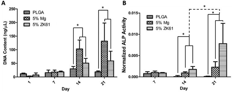
Figure 8.

In vitro biocompatibility. Mouse pre-osteoblastic (MC3T3-E1) cell performance and osteogenic differentiation on PLGA, PLGA-Mg, and PLGA-ZK61 was assessed through DNA quantification and ALP expression. (A) Cellular proliferation was analyzed using Picogreen dsDNA assay. Increased cell proliferation for Mg and ZK61 samples was observed at days 14 and 21, with little change in DNA concentration in PLGA group throughout the study. (B) ALP expression was analyzed using an ALP assay kit and normalized to DNA concentration. Increased ALP expression was seen between PLGA and the composite groups at days 14 and 21. Significant increase in ALP expression between day 14 and 21 was also seen for the PLGA-Mg and PLGA-ZK61 samples (n=3* = p <0.05).
