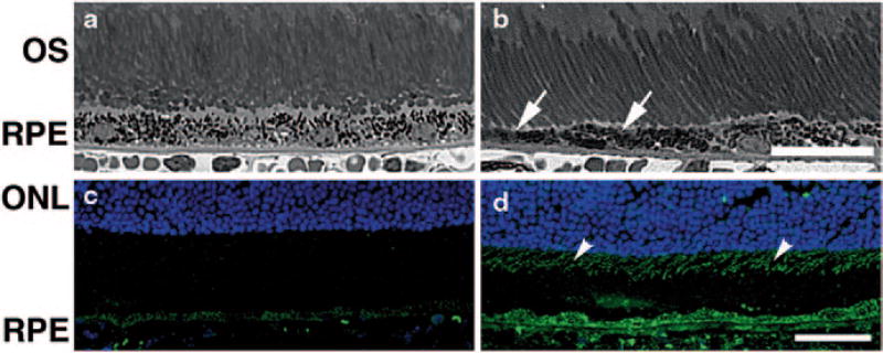
Fig. 81.3.
Degeneration in the neural retina of young adult DJ-1 KO mouse. Plastic sections of both control (a) and DJ-1 KO (b) retinas stained with toluidine blue highlighted the RPE and photoreceptor cells. The RPE of the DJ-1 KO displayed thinning (b, arrows). Cryosections of retinas were also labeled with 8-oxoG (c, d) to detect DNA oxidation. Analysis showed that 8-oxoG immunoreactivity is significantly increased in the photoreceptor inner segments (arrowheads) and the RPE cells of DJ-1 KO (d) when compared to the control (c) retinas. ONL outer nuclear layer, OS photoreceptor outer segments, RPE retinal pigment epithelium, Bar a, b = 25 µm and c, d = 40 µm

