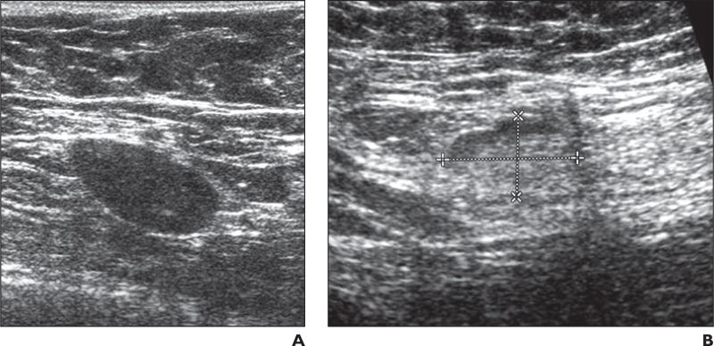
Fig. 4. Two patients with fatty hilum within lymph node.
A, 50-year-old woman with biopsy-proven nodal metastases from invasive breast cancer. Ultrasound examination performed after patient received chemotherapy revealed lymph node that remained abnormal with persistent loss of fatty hilum in node (type VI). Final pathologic analysis confirmed 0.7-cm metastasis in this 2.4-cm lymph node.
B, 57-year-old woman with biopsyproven metastatic node from invasive breast cancer. Ultrasound examination performed after patient received chemotherapy revealed normal-appearing lymph node with visible fatty hilum suggesting no residual disease. Dashed lines show long and short axis of node. Final pathologic analysis revealed 0.3-cm metastasis in this 1.8-cm lymph node.

