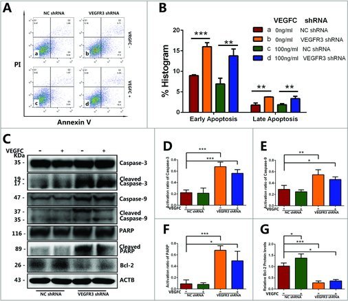
Figure 6.

The effects of VEGFC/VEGFR3 on the early and late apoptosis of GC-1 cells. (A) Flow cytometry demonstrated the influence of VEGFR3 knockdown and VEGFC treatment on the early apoptosis (Q3) and late apoptosis (Q2) of GC-1 cells. The existence of Annexin V (X-axis) and the nuclear staining of PI (Y-axis) by flow cytometry were shown. (B) Annexin V/PI flow cytometry analysis showed that VEGFR3 knockdown led to increases in apoptosis in GC-1 cells compared to the negative control shRNA. (C-G) Western blotting showed the transcript levels of apoptosis protein in GC-1 cells treated with VEGFR3 shRNA or negative control shRNA. The activation ratio of Caspase-3 protein (D), Caspase-9 protein (E), and PARP (F) and Bcl-2 protein (G) was showed in the form of histogram. Values in (A-G) were represented as mean ± SD from three independent experiments. (*P<0.05, **P<0.01, ***P<0.001).
