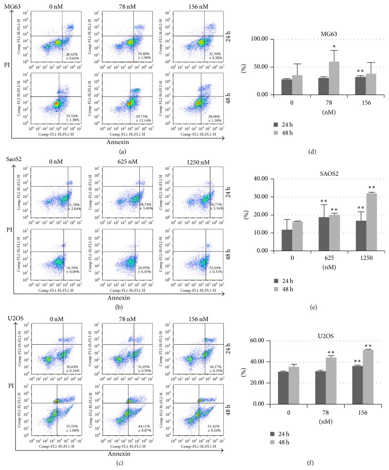
Figure 3.
The effect of Rg3 on apoptosis and necrosis of MG63, SaOS-2, and U-2OS cell lines evaluated by flow cytometry assay. The cells were double-stained with FITC-Annexin V and propidium iodide (PI). Mean values of the percentage of apoptotic and necrotic cells from four independent experiments ± SD are presented. Percentage of apoptotic cells was the sum of percentage early apoptotic (LR) and late apoptotic cells (UR). ∗p < 0.05; ∗∗p < 0.001.

