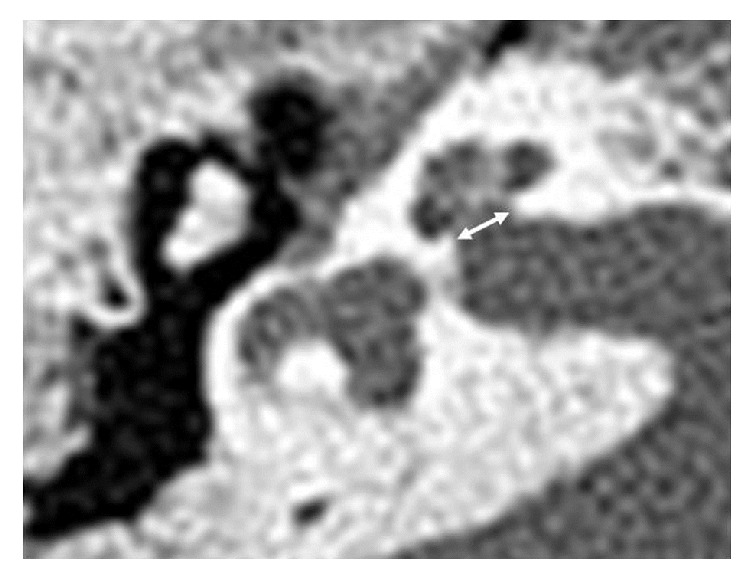
Figure 1.

Axial slices of temporal bone computed tomography for the measurement of bony cochlear nerve canal (BCNC) in normal bony cochlear nerve canal. The width was measured by the distance between the inner margins of bony walls at midportion, at the fundus level of cochlear nerve in internal auditory canal.
