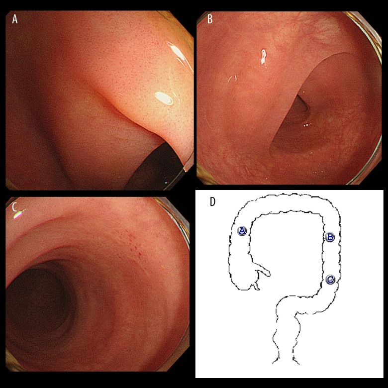
Figure 2.
Endoscopic finding of the colon mucosa. (A–C) The endoscopic images of the ascending colon, the descending colon, and the sigmoid colon, respectively. There was enteritis with mild edema affecting the whole colon mucosa. However, ulcerative lesions were not observed at any site. The findings were compatible with enterocolitis associated with nivolumab. (D) Illustrates the sites of the colon where A–C were taken.

