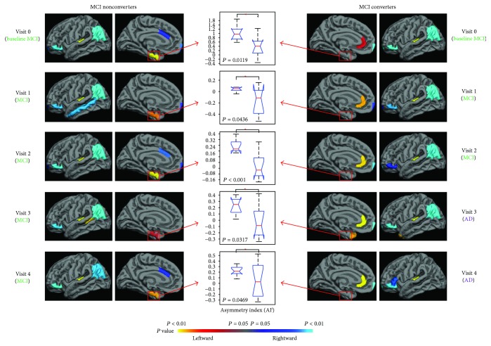
Figure 1.
Hemispheric asymmetry analysis demonstrated different patterns in the medial side of the brain between the MCI nonconverters and converters at all time points. The MCI nonconverters showed leftward lateralization in the entorhinal cortex, while the converters showed rightward lateralization in the rostral cingulate gyrus. Significant between-group difference of the asymmetry index AIj was observed in these two regions with t-test analysis (middle column).

