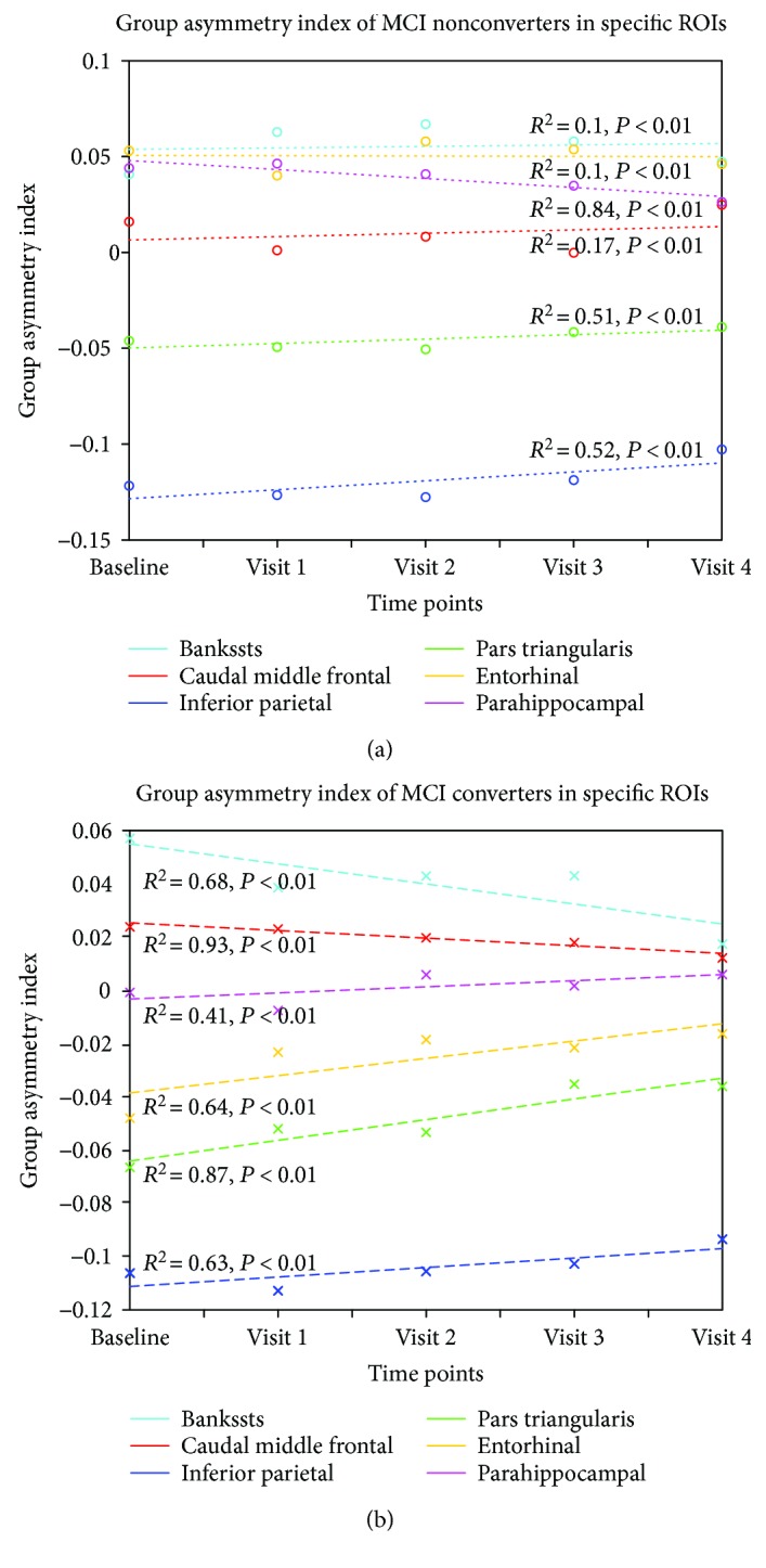
Figure 3.

Regression of group asymmetry indices for MCI nonconverters and converters in five selected ROIs, including the banks of the superior temporal sulcus (cyan), caudal middle frontal gyrus (red), inferior parietal lobule (blue), pars triangularis (green), entorhinal cortex (yellow), and parahippocampal gyrus (pink). Group asymmetry indices decreased more notably to 0 in MCI converters from baseline to follow-up visits compared to nonconverters. Bankssts: banks of the superior temporal sulcus.
