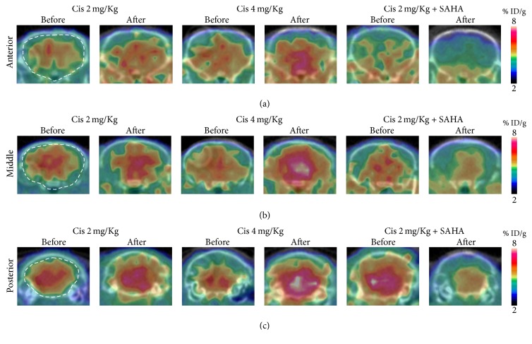
Figure 2.
Coronal brain image of [18F]FAHA before and after drug administration. [18F]FAHA PET/CT images summed over the last 10 min of the study (20–30 min after [18F]FAHA injection). (a) Anterior, (b) middle, and (c) posterior part of the brain. Brain [18F]FAHA radioactivity was significantly increased after cisplatin administration.

