Figure 3.
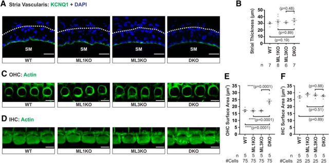
There is an anatomical defect in OHCs but not in the stria vascularis from cochleae lacking both mucolipin 3 and 1. A, Immunoreactivity of potassium channel KCNQ1 in the stria vascularis shows that there is no mislocalization of this channel in DKO cochlea compared with WT, ML1KO, and ML3KO animals. SM, Scala media. B, Quantification of strial thickness indicates that there is no strial degeneration in DKO cochlea. C, D, Actin labeling reveals that OHCs (C), but not IHCs (D), from DKO cochlea are larger than those of control cochleae. E, F, Quantification of HC apical surface area, measured just under the cuticular plate of HCs from middle turn, confirms that DKO OHCs are larger than the controls. Samples were from 4- to 4.5-month-old animals. Scale bars: A, 20 μm; C, D, 5 μm. ****p ≤ 0.0001.

