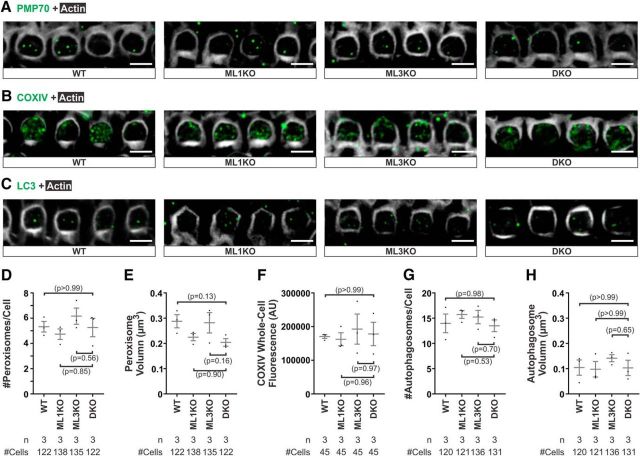
Figure 6.
Enlarged lysosomes in OHCs lacking mucolipin 3 and 1 do not cause defects in lysosomal degradation of damaged organelles. A–C, Whole-mount cochleae from WT, ML1KO, ML3KO, and DKO adult (∼P120) animals using different organelle markers: peroxisome (PMP70, A), mitochondria (COXIV, B), and autophagosome (LC3, C). There is no apparent defect in distributions and numbers of peroxisomes (A), mitochondria (B), and autophagosomes (C). Representative images were reconstructed from a projection of 8 confocal optical sections spanning 0.88 μm under the cuticular plate. D, E, The number of peroxisomes per OHC and peroxisomal volume, indicated by PMP70 immunoreactivity, from DKO OHCs are similar to those from controls. F, Whole-cell fluorescence of COXIV did not differ between DKO and control OHCs. G, H, The number of autophagosomes per OHC and autophagosomal volume, indicated by LC3 immunoreactivity, from DKO OHCs are similar to controls. Scale bars, 5 μm.

