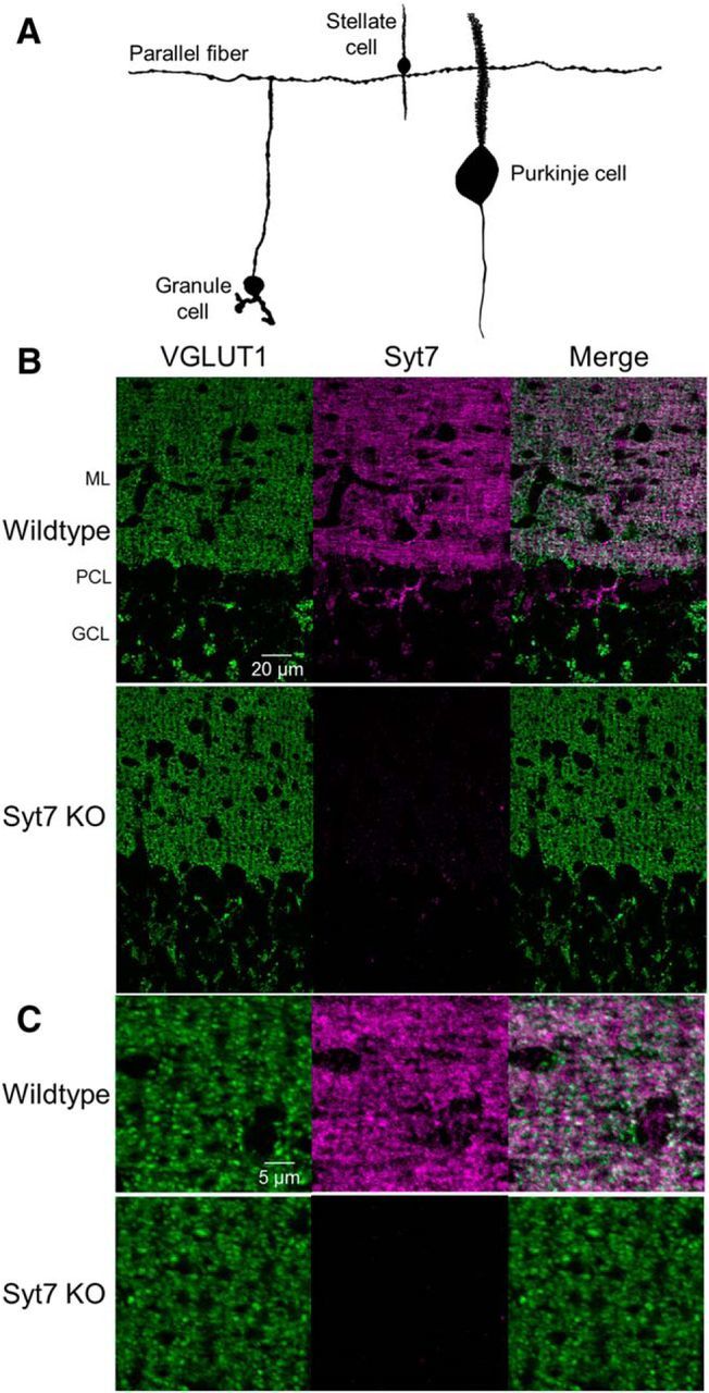
Figure 1.

Immunohistochemical localization of Syt7 in the cerebellar cortex. A, Parallel fibers originate from granule cells and extend laterally in transverse sections, forming synapses onto stellate and PCs in the cerebellar cortex (vermal lobule VI). The SC and PC dendrites are oriented perpendicular to the surface of the slice. B, Transverse sections of cerebellar cortex immunolabeled for the presynaptic marker VGLUT1 (green) and Syt7 (magenta) in a wild-type (top) and Syt7 KO (bottom). ML, Molecular layer; PCL, Purkinje cell layer; GCL, granule cell layer. C, Expanded view of the molecular layer for the images in B.
