Figure 5.
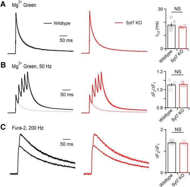
Presynaptic Ca2+ signaling is unaltered in Syt7 KO animals. A, Parallel fibers were loaded with the low affinity Ca2+ indicator Mg-Green-AM (Kd = 7 μm for Ca2+), and stimulated. Ca2+-dependent presynaptic fluorescence changes are shown for average wild-type (black) and Syt7 KO animal (red). The half decay time was measured and summarized (right; wild-type: n = 6, Syt7 KO: n = 6). B, Same as in A, but for brief stimulus trains (5 stimuli at 50 Hz). Averages are shown for wild-type and Syt7 KOs (left) with single stimuli overlaid. The ratio between the amplitude of the first and second transient in the train are summarized (right; wild-type: n = 7, Syt7 KO: n = 6). C, Parallel fibers were labeled with the high affinity Ca2+ indicator Fura-2-AM (Kd = 0.2 μm) and responses were evoked by one and two stimuli. Averages are shown for a wild-type (black) and Syt7 KO (red). The ratio of the amplitudes of the responses evoked by two stimuli and one stimulus provides a measure of the magnitude of presynaptic Ca2+ entry, and is summarized (right; wild-type: n = 9, Syt7 KO: n = 10). Traces are normalized to peak values evoked by a single stimulus. Data are mean ± SEM.

