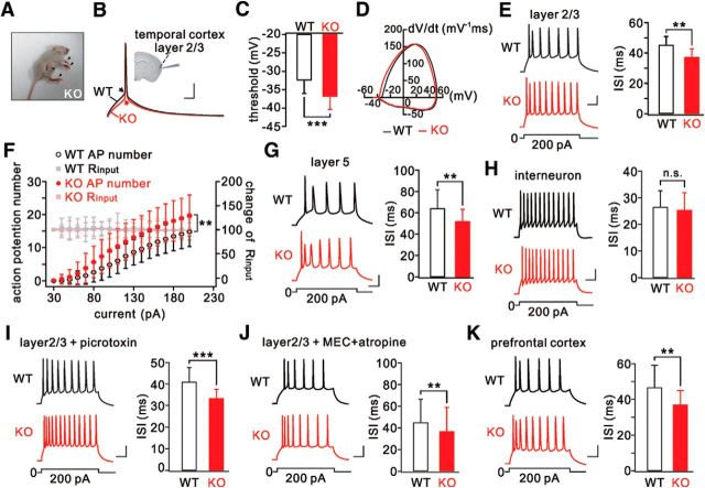
Figure 1.
Hyperexcitability in temporal cortical pyramidal neurons from LGI1−/− (KO) mice. A, Arrowheads indicate tonic limb extension after spontaneous generalized seizures in a KO mouse (P16). B, Action potentials induced by injecting a depolarizing current (50 ms) in WT and KO pyramidal neurons. Arrows indicate initiation of action potentials. Inset, Location of recordings in the temporal cortex. C, Summary of voltage thresholds (WT: −32.3 ± 3.8 mV, n = 23; KO: −36.7 ± 3.4 mV, n = 25). D, Phase-plane plots (Vm vs dV/dt) for single action potentials induced in WT and KO neurons. The voltage threshold for the action potential in the KO neuron shifted to a more hyperpolarized level. E, Sample spikes from WT and KO neurons in response to a 300 ms current injection (200 pA) and summary of ISIs. F, Input–output curves for WT (n = 34) and KO (n = 22) neurons showing numbers of action potentials as a function of injected currents lasting 800 ms. Input resistances (Rinput) were monitored during the course of experiments. All Rinput values were normalized to the first point, and the percentages changes are on the y axis on the right. G, Spikes of WT and KO layer 5 neurons in response to current injection. H, Spikes and ISIs in WT and KO parvalbumin-positive interneurons. I, Spikes in WT and KO layer 2/3 neurons treated with picrotoxin (100 μm). J, Spikes and ISIs in WT and KO layer 2/3 neurons treated with mecamylamine (MEC; 10 μm) and atropine (1 μm). K, Spikes and ISIs in WT and KO prefrontal cortex. Calibration: 20 mV, 50 ms. **p < 0.01, ***p < 0.001.

