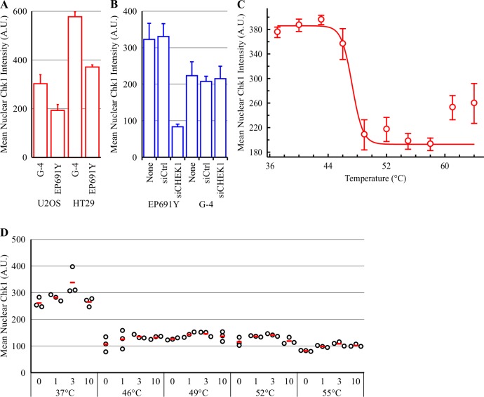
Fig 3. V158411 does not thermostabilize Chk1 in intact cells heated by the PBS immersion method.
(A) Mean nuclear Chk1 intensity was determined in U2OS and HT29 cells following immunofluorescent staining with the anti-Chk1 antibodies G-4 and EP691Y and high content imaging. Mean of 3 independent wells ± SD. (B) HT29 cells were transiently transfected with control (siCtrl) or CHEK1 smart pool siRNA (siCHEK1) for 48 hours. Mean nuclear Chk1 intensity was determined by immunofluorescent staining with G-4 or EP691Y antibodies and high content imaging. (C) Thermal response profile of Chk1 in HT29 cells detected by immunofluorescent imaging with the EP691Y antibody following immersion in PBS pre-heated to the indicated temperature. Values are the mean of four wells ± SD. (D) HT29 cells were treated with 0–10 μM V158411 for 30 minutes before being heated by immersion in PBS pre-heated to the indicated temperature for 3 minutes followed by room temperature for 5 minutes. Nuclear Chk1 intensity was determined by immunofluorescent staining using the EP691Y antibody and high content imaging. Red line, mean.

