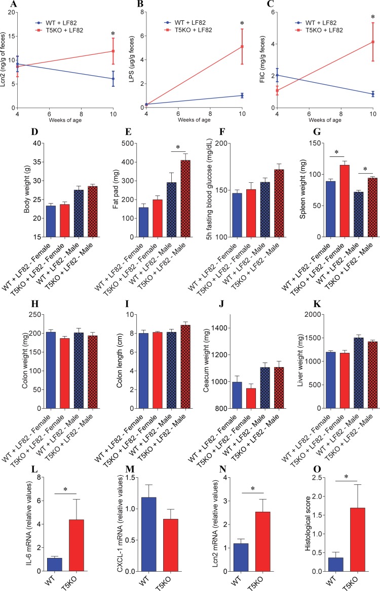
Fig 5. AIEC colonization induces modest increase in low-grade inflammation and adiposity in ASF-T5KO mice.
Four-week old offspring of ASF-colonized WT and T5KO mice were removed from the isolator, placed in isolated ventilated cages and inoculated with AIEC reference strain LF82 placed in drinking water for two weeks, followed by return to autoclaved water. At 12 weeks of age, animals were removed from this isolator and euthanized. A. Fecal Lcn2 levels at week 4 and week 10 of age. B. Fecal lipopolysaccharide (LPS) levels at week 4 and week 10 of age. C. Fecal flagellin (FliC) levels at week 4 and week 10 of age. D. Final body weight. E. Fat pad weight. F. 5 h fasting blood glucose concentration. G. Spleen weight. H. Colon weight. I. Colon length. J. Caecum weight. K. Liver weight. L-N. Colonic pro-inflammatory cytokine-encoding genes were quantified by qRT-PCR (L, IL-6; M, CXCL-1; N, Lcn2). O. Hematoxylin & eosin staining was performed on colonic sections and used for the determination of histopathological scores. Data are the means +/- S.E.M. (n = 3–6). Significance was determined using t-test (* indicates p<0.05). Data are the means +/- S.E.M. (n = 3–6). Data in A, B and C combine both male and female animals. Significance was determined using t-test (* indicates p<0.05).

